3D Anatomy Atlas
Rotate an organ, peel it apart, slice it at any level next to a real CT or MRI section of the same level, and quiz yourself on every structure. Built from open, credited anatomy data. Runs in your browser. Sign in to open an atlas: you get one free atlas visit, and a subscription opens all of them.
 Surgery · Radiology · GI
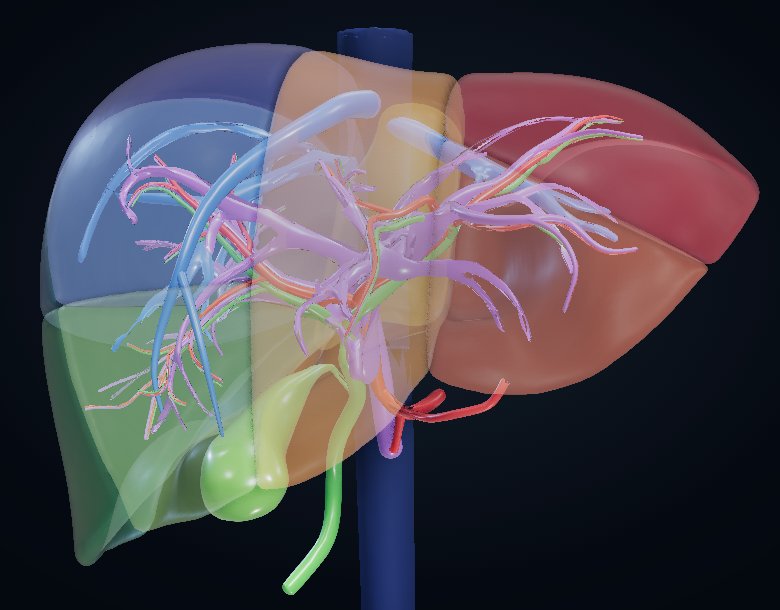
Liver
Couinaud segments I–VIII with the portal, arterial, hepatic-vein and biliary trees, a level-matched axial CT and a resection planner.
- • Slice the 3D model alongside a real CT
- • Brisbane 2000 resections with remnant %
 Surgery · Radiology
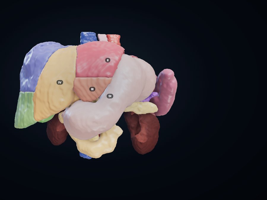
Upper abdomen
Built from a real CT: liver segments, stomach, duodenum, pancreas, spleen, gallbladder, portal–splenic vein, aorta and IVC in true relations.
- • Axial and coronal CT of the same person
- • Real CT anatomy
 Medicine · Cardiology · Anaesthesia
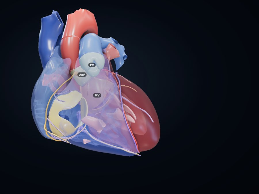
Heart
Four chambers and septum, all four valves, papillary muscles, LM, LAD, diagonals, marginals, RCA and PDA, and the cardiac veins.
- • Cardiac CT matched by level
- • Coronary territories with ECG leads
- • Glass mode shows the valves
 Medicine · Pulmonology · Thoracic surgery
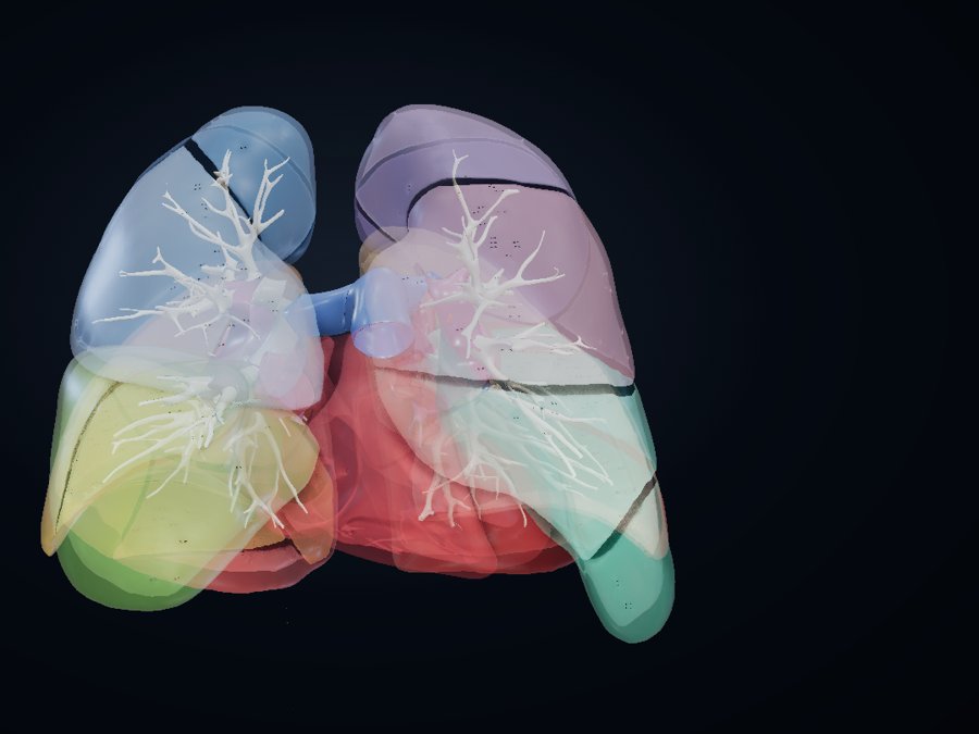
Lungs
All 20 bronchopulmonary segments, the bronchial tree to segmental bronchi, hilum and pulmonary vessels.
- • Lung-window CT with the lobes
- • Segments numbered R1–R10, L1–L10
 Neurology · Neurosurgery · Psychiatry
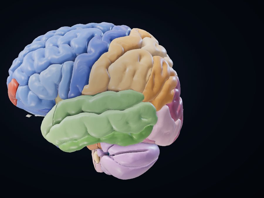
Brain & brainstem
Every gyrus by lobe, basal ganglia and thalamic nuclei, hippocampus and amygdala, midbrain, pons, medulla, cerebellum and ventricles.
- • T1 and T2 MRI: axial, coronal, sagittal
- • Allen Human Brain Atlas structures
 Neurology · Neurosurgery · Neuroradiology
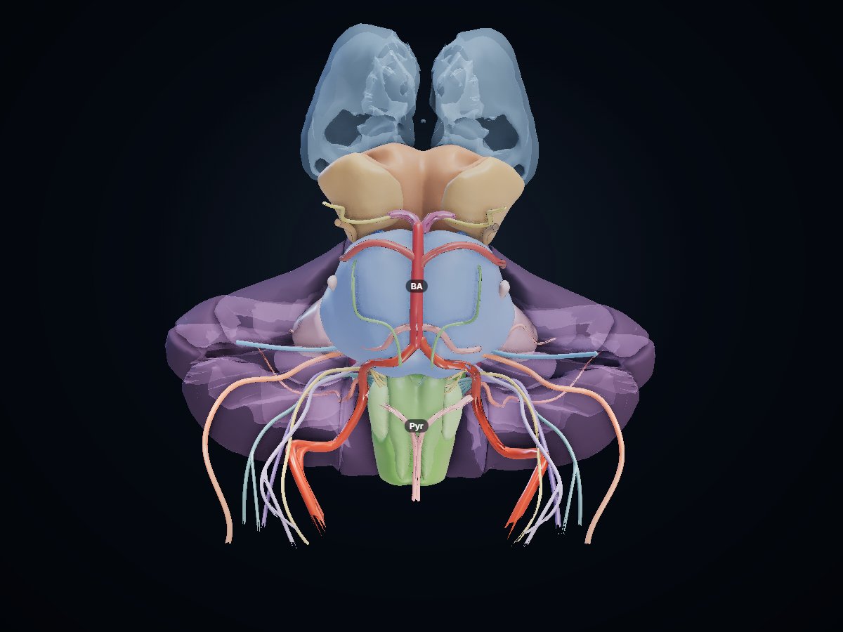
Brainstem
Midbrain, pons and medulla with colliculi, peduncles, olives and pyramids, red nucleus and substantia nigra, cranial nerves III–XII at their exits, and the vertebrobasilar arteries.
- • Axial and sagittal MRI matched by level
- • Brainstem syndromes in the notes
 Urology · Nephrology · Radiology
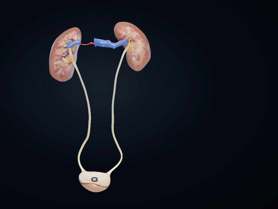
Kidney, ureter, bladder
Capsule, cortex, renal columns, pyramids and papillae, calyces, renal pelvis, ureters, trigone and renal vessels.
- • CT from kidneys to bladder
- • Glass kidney shows the medulla
- • Stone impaction sites
 Urology
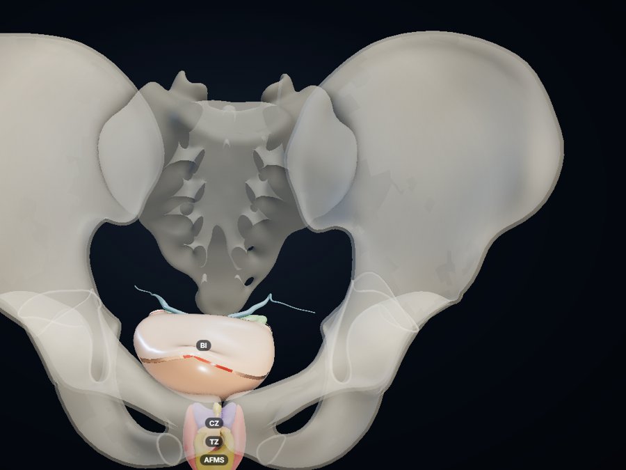
Male pelvis
McNeal prostate zones, seminal vesicles, vasa, ejaculatory ducts, verumontanum, bladder and bony pelvis.
- • CT and prostate zonal MRI
- • Peripheral, transition, central zones
- • TURP landmarks
 OBGY
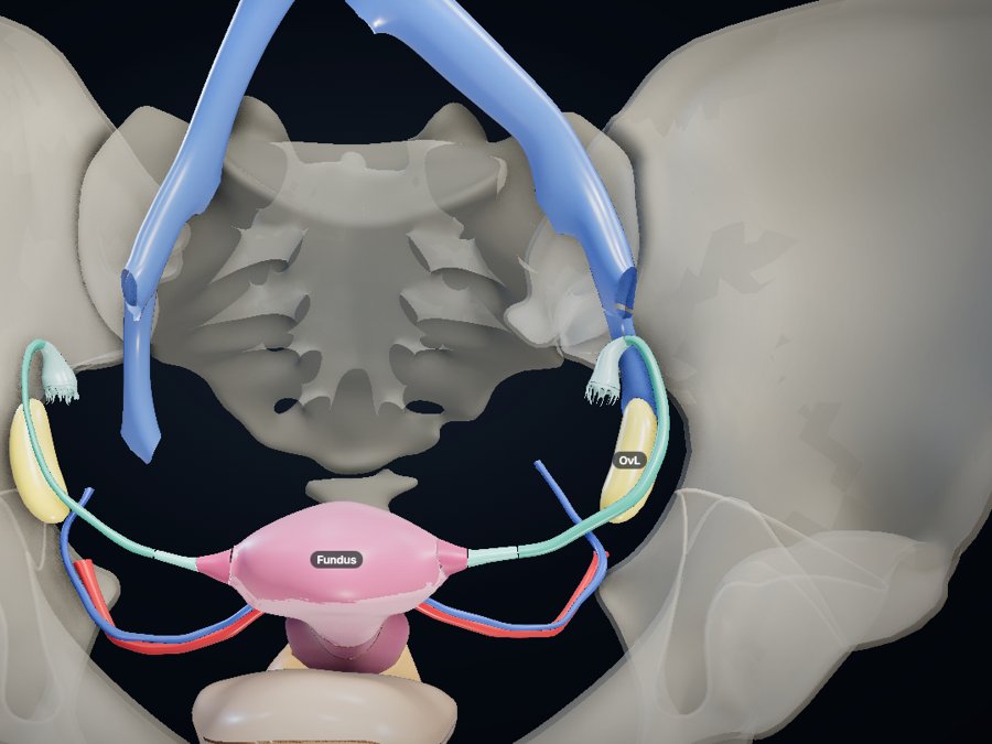
OBGYFemale pelvis
Fundus, body, lower segment, cervix and os, tubes by segment, ovaries, uterine vessels, bladder and bony pelvis.
- • CT and pelvic MRI with uterus and ovary
- • Uterine artery and ureter relation
- • Ectopic pregnancy sites
 Ophthalmology
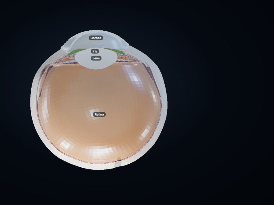
Eye
Sclera, cornea, uvea, retina with macula, fovea and disc, lens and zonules, trabecular meshwork and Schlemm’s canal.
- • Horizontal section of the globe
- • Drainage angle in 3D
 ENT · Anaesthesia · Dental
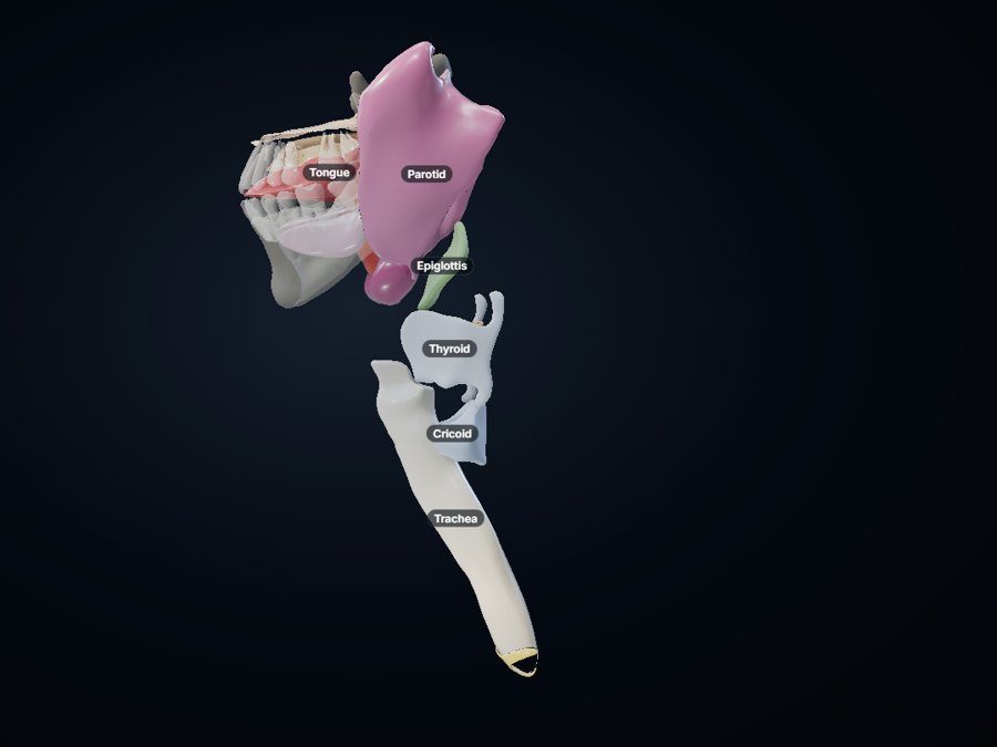
Head & neck
Laryngeal cartilages, trachea and carina, tongue, palate, teeth, parotid, submandibular and sublingual glands, tonsils.
- • Neck CT with larynx and glands labelled
- • Explode the larynx
- • Airway landmarks for intubation
 Neurosurgery · ENT · Radiology
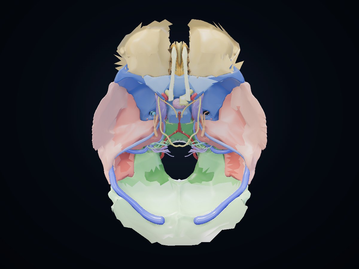
Skull base
Anterior, middle and posterior cranial fossae bone by bone, every major foramen ringed with what passes through it, cranial nerves I–XII, carotid, jugular vein and dural sinuses.
- • Axial and coronal CT
- • Foramina with their contents
 Ophthalmology · Neurology
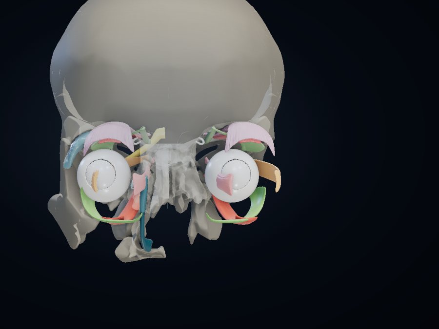
Orbit
Six extraocular muscles and levator, trochlea, optic nerve, ciliary ganglion, globe and the whole lacrimal drainage system inside the orbital bones.
- • Coronal orbit MRI
- • Explode the muscles
- • Cranial nerve palsy notes
 ENT · Radiology
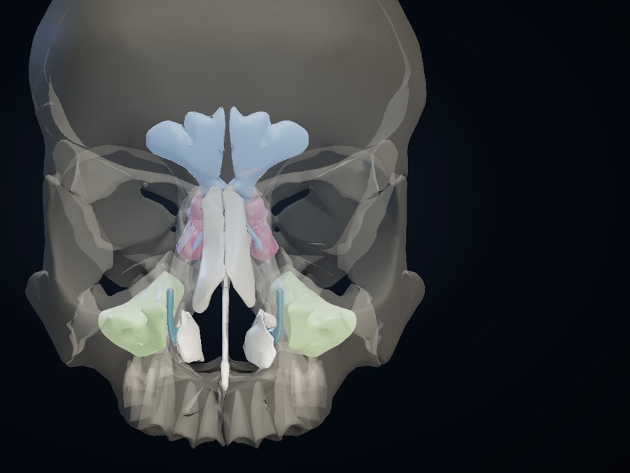
Nose & sinuses
Frontal, maxillary and sphenoid sinuses, anterior, middle and posterior ethmoid cells, ethmoidal bulla and nasolacrimal ducts in a translucent skull.
- • Sinus CT
- • Drainage pathways in the notes
- • Explode the sinuses
 ENT · Audiology
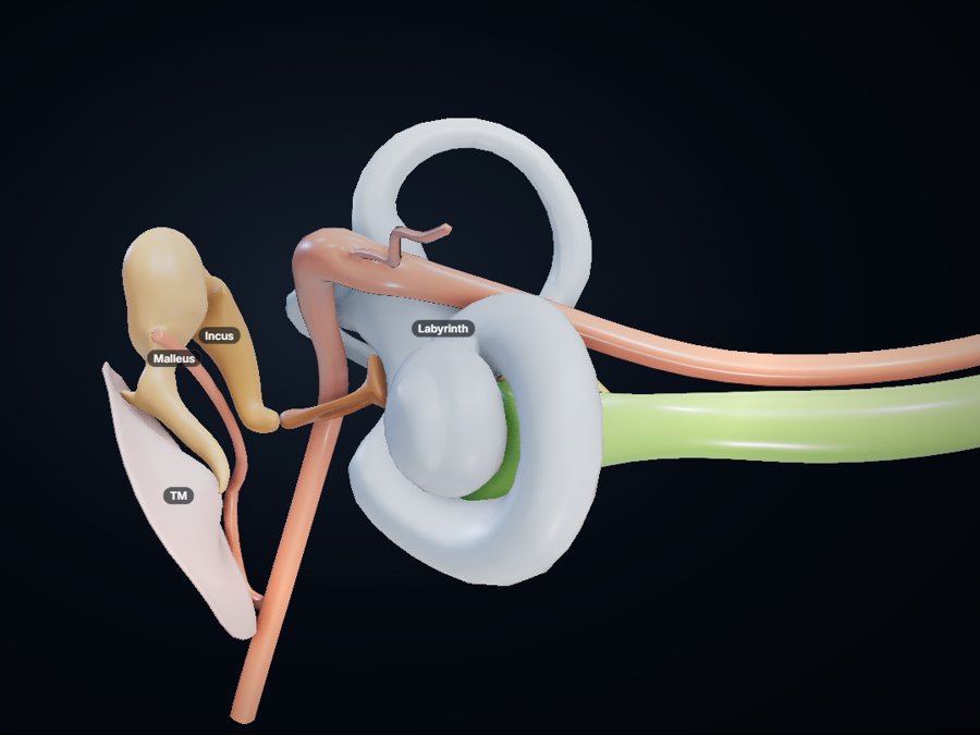
Ear
Tympanic membrane, malleus, incus and stapes, the bony labyrinth, and the cochlear, vestibular and facial nerves.
- • Temporal-bone CT, axial and coronal
- • Facial nerve course
- • Glass labyrinth
 Orthopaedics · Plastic surgery
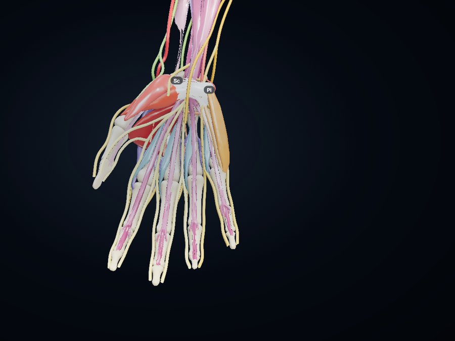
Hand
All 27 bones, thenar and hypothenar muscles, interossei and lumbricals, tendons, palmar arches, and the median, ulnar and radial nerves.
- • Hand CT cross-sections
- • Carpal-tunnel cross-section
- • Nerve-injury signs in the notes
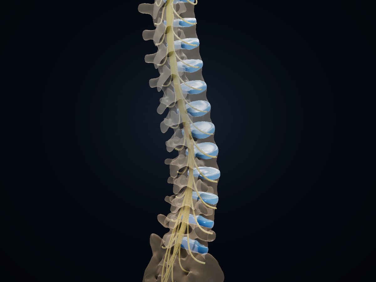 Orthopaedics · Neurosurgery · Anaesthesia · Radiology
Spine
Every vertebra from atlas to L5, sacrum, intervertebral discs, spinal cord with conus and filum, nerve roots and cauda equina, and vertebral arteries.
- • Vertebra-matched CT and lumbar MRI
- • Cord segments vs vertebral levels
- • Disc prolapse: which root?
For teaching only, not for planning a patient’s treatment. 3D sources: Human Reference Atlas 3D Reference Object Library (HuBMAP / CNS Indiana University; Allen Human Brain Atlas structures), CC BY 4.0; BodyParts3D, © The Database Center for Life Science, CC BY 4.0; Z-Anatomy (CC BY-SA); Sketchfab models by OSU.Multimedia (CC BY) and anatomary (CC BY-NC); TotalSegmentator dataset (Wasserthal et al., Radiology: AI 2023), CC BY 4.0. Full credits inside each atlas.